Fusion of Imaging Data (CT-MR, CT-CT, MR-MR)
Overview
Fusion transform between MR sequence and CT sequence as reference model can be calculated with known statistical method (optimization based on overlapping image informations: Maes F, Vandermeulen D, Suetens P. Comparative evaluation of multiresolution optimization strategies for multimodality image registration by maximization of mutual information. Medical Image Analysis 1999; 3(4):373-386.). The surgical planning program takes samples from both fusion volume and reference volume with predefined, initial voxel steps (in x-y-z). The calculations are sped up by noise removal applied for reference images. The calculations use the known Powell numerical optimization in 3D (Press WH, Flannery BP, Teukolsky SA, Vetterling WT. Numerical Recipes in C, second edition. Cambridge, Cambridge University Press, 1992:394-420.). The program sets identical voxel size for reference and fusion sequences by initial interpolation of fusion sequence.
The program is able to restrict fusion registration to arbitrarily selected subvolume (VOI,”Volume of interest”), as target volume. Using this method, one can select the most relevant, overlapping and noise-free part of volumetric models where the computation is executed by high accuracy. This method is especially important for short T2 sequences and fusion models with limited overlap. Accurate registration of fusion sequence needs acceptable fit of overlapping subvolumes, relevant assignment.
The fusion algorithm defines the linkage between reference and fusion models. The panel “CT-MR Fusion” controls all of details of fusion computations and, in case of acceptable result, it stores the fusion transform. This transform with the coordinates of subvolumes (VOI) is copied into planning panel and is stored in archive study file (.vts) together with the surgical plan. After turning off VOI frames the fusion transform is converted for the whole volume. Transparent display helps visualizing fusion (reference image as foreground and registered fusion image as background).
Selection of Targeting Subvolumes (VOI)
The initial parameters of fusion target subvolume can be set on reference series by modifying the size and 3D position with the help of axial and frontal views (Image 7-8, on left).). This The first step defines the size and 3D position of target subvolume on fusioned imaging data (Image 7-8, on right). Only the VOI position (size not) can be moved on fusioned background; the goal is to find the best overlap between reference and fusioned subvolumes. Using this method, the user is able targeting the anatomical area with registration optimum.
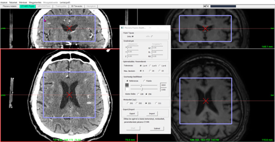
Image 7. Fusion subvolumes (VOI) for MR T1 (VOI) with CT reference.
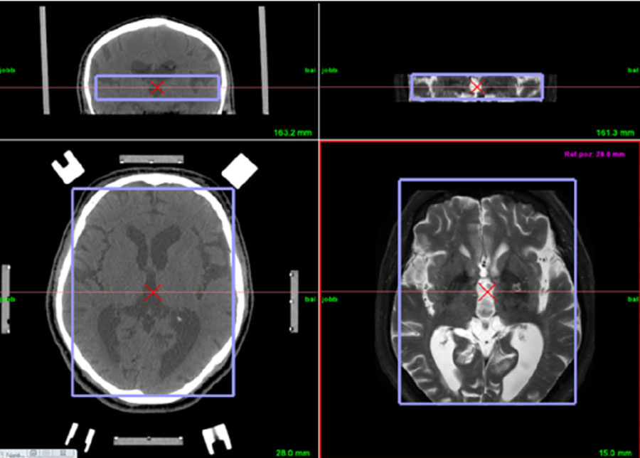
Image 8. MR T2 sequences initialized in CT reference view. VOI adjusted according to the short T2 sequence.
Control Settings/Optimization
After reading image sequences the program automatically fills up the control window with initial parameters or, in case of archived (.vts) file with the results of earlier calculations (Fusion Settings). In case of new fusion calculation the numerical parameters and grayscale limits can be modified both for references and fusion sequences. According to fusion algorithm the rate of voxel sampling (xyz: 331 332 221 111) can be used to speed up the calculations. The user can set the error tolerance (1e-4, 1e-5, 1e-6) and the number of iteration loops (5, 10, 15). After some practice, the fusion can be easily parameterized to frequent diagnostic settings and surgical interventions and the optimized solution can be found under the conditions of local diagnostic environment. By the help of export/import functions the projection transformation can be archived and recalled. During import the actual registration transform can be replaced or modified (in case of modifying the existing transformation is multiplied, continues to transform).
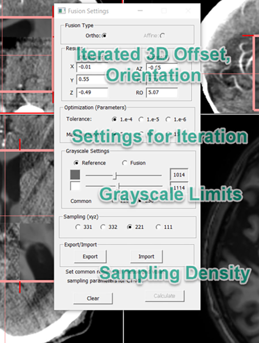
Image 9. Panel for fusion calculations. Right lower button starts the computation (after locking the actual VOI settings). The resulted offset and orientation values between the MR data and reference CT can be seen on the upper part of dialog window. In case of inaccurate result the actual offset and rotation data can be used as initial values for a repeated registration calculation. Manual adjustment is possible for axial, frontal, sagittal offset values and orientation angles.
Displaying Results of Image Fusion
After some seconds, depending on the hardware configuration and parameterization, CranioPass finishes the calculation and according to the result the fusioned image sequence will show up as background image in CT views.
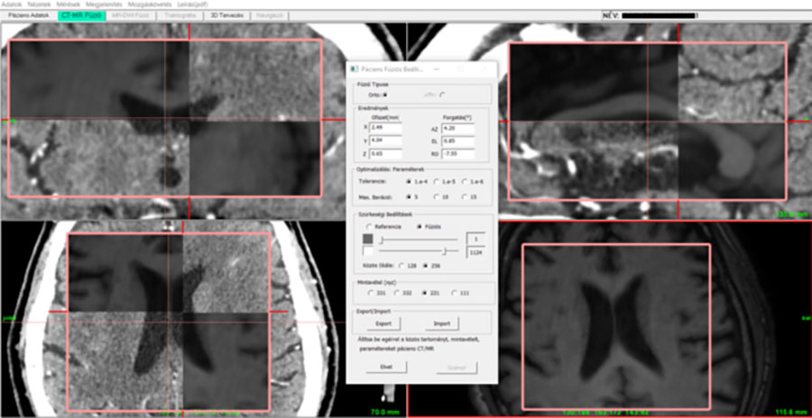
Image 10. Display of fusioned views with reference sequence reformatted into axial, coronar and sagital directions. In case of false result (inaccurate fusion overlap) the registration can be repeated with new parameters or modified initial values (starting new optimization). After that the fusion and reference views can be compared with new sampling.
6DOF Modification of Fusion Parameters
The fusion offset/orientation values (6DOF) can be adjusted in control window without numerical optimization just by setting new value for any component of xyz offsets or 3 orientation angles. Setting new 6DOF value automatically updates fusion sequence (by interpolated resampling) and visualizes the new fusioned background in all three (axial, sagittal, frontal) views. This manual method is possible only in locked views. Reference sequences with modified fusion background can be investigated in transparent or non-transparent mode.
Lock/Unlock of Fusion Results
Locking/unlocking fusion results is possible by double click in axial view (“Lock/Unlock”). If locked fusion is cleared, the program initiates request to user if he wish to keep registration or not.
Import/Replacement of Fusion Sequence and PostOp Import
The actual fusion sequence can be overwritten with an imported series. The program asks for if the current fusion parameters (offset, angles) should be replaced or not. If the user keeps the results the new sequence overwrites the old one but it is reformatted with existing fusion transform. Using this method the modified version of the actual fusion sequence (with added image information, like MR with calculated tractographic data) can be inserted with valid registration into surgical plan. The fusion transform can be calculated with high precision using the original sequence and, in a following step, the new sequence with added functional elements can be inserted with the original transformation. Similar task comes up after surgery investigations (PostOp) where the PostOp imaging data should be registered to the original, reference volume in such a way that the planning data (target coordinates etc), the positions stored in the original planning srudy (.vts archive) should remain constant. After importing PostOp CT or MR, the planning software re-registers fusion sequence and the 3D planning positions can be displayed.
CranioPass CranioPassCranioPassCranioPass
CranioPassCranioPassCranioPassCranioPassCranioPassCranioPassCranioPassCranioPassCranioPassCranioPassCranioPassCranioPassCranioPassCranioPassCranioPassCranioPassCranioPassCranioPassCranioPassCranioPassCranioPassCranioPassCranioPassCranioPassCranioPassCranioPassCranioPassCranioPassCranioPassCranioPassCranioPassCranioPassCranioPassCranioPassCranioPassCranioPassCranioPassCranioPass